No price shown online — add this to your list and we'll quote you based on the quantity you need.
Prefer to talk it through? Contact us or WhatsApp directly.
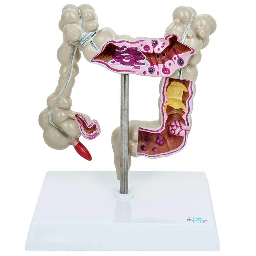
The Human Colon Model is an anatomical representation of the human colon that aims to mimic its structure and/or function for research and educational purposes. These models can be used to study the colon's anatomy, the large intestines shows the anatomy as well as several common diseases and conditions.
The colon is cross-sectioned to show colitis, appendicitis, adhesions, diverticulitis, diverticulosis, bacterial infection, cancer, spastic colon, and polyps. Crohn’s disease is shown in an area of the small intestines. The appendix and rectum are also shown.
A cross-section of the colon, also known as the large intestine, reveals its internal structure, which includes the mucosa, submucosa, muscularis externa, and serosa. The colon is a muscular tube that extends from the cecum to the rectum and is characterized by its folds, haustra (pouches), and epiploic appendages (fatty tags).
Understanding the cross-sectional anatomy of the colon is crucial for diagnosing and treating various conditions, including inflammatory bowel disease, diverticulitis, and colorectal cancer.
Mucosa: The innermost layer, containing the lining epithelium, lamina propria, and muscularis mucosae.
Submucosa: A layer of connective tissue beneath the mucosa, containing blood vessels, nerves, and lymphatic vessels.
Muscularis Externa: A muscular layer responsible for peristalsis (movement of contents through the colon), composed of inner circular and outer longitudinal muscle layers.
Serosa: The outermost layer, a thin membrane that covers the colon.
Key features visible in a cross-section:
Educational Tool: This model is intended for educational use, particularly in biology and medical training, providing a hands-on learning experience with a detailed anatomical representation.